Beautiful embryo photos

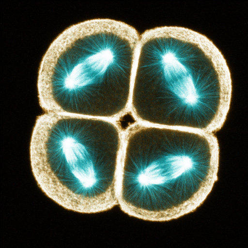
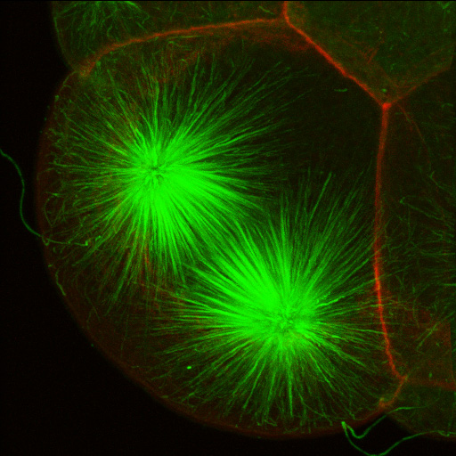
There are some gorgeous embryos out there. As an evolutionary developmental biologist I can’t help but take a little detour from jellies to post some of these stunning works. Images and subtext are from The Center for Cell Dynamics website (except those things in italics and parenthesis, which are from me).

Single confocal section of an oocyte of the nemertean Cerebratulus in metaphase of first meiosis. Microtubules are orange, actin is blue; one pole of the meiotic spindle is anchored to the cortex, and the other, deep in the cytoplasm, exhibits long astral rays. (George von Dassow)
Four-cell embryo of the protobranch bivalve Acila castrenis (clam) fixed and stained for F-actin using phalloidin; projection of one-micron sections (George von Dassow)
Four-cell embryo of the protobranch bivalve clam Acila castrenis  fixed and stained for F-actin using phalloidin; projection of one-micron sections (George von Dassow)
ingle confocal section through four cells of an eight-cell sand dollar embryo fixed and stained with anti-tubulin (green) and phalloidin (red). Vegetal cells are dividing asymmetrically to make micromeres and macromeres (George von Dassow)
Single confocal section through four cells of an eight-cell sand dollar embryo fixed and stained with anti-tubulin (green) and phalloidin (red). Vegetal cells are dividing asymmetrically to make micromeres and macromeres (George von Dassow)
Single confocal section of a 2-cell embryo of the nemertean Cerebratulus. Microtubules are bronze and DNA is blue. (George von Dassow)
Mitotic spindles in the four animal blastomeres of an eight-cell purple urchin embryo, fixed and stained with anti-tubulin (blue) and phalloidin (orange); projected from serial half-micron sections (George von Dassow)
Mitotic spindle at approximately metaphase in one cell in a 16-cell embryo of the clam Acila. Microtubules in green, actin in red. Projection from serial 0.5-micron confocal sections. (George von Dassow)
Advertisement

Leave a Reply

Fill in your details below or click an icon to log in:

WordPress.com Logo

You are commenting using your WordPress.com account. Log Out /  Change )

Facebook photo

You are commenting using your Facebook account. Log Out /  Change )

Connecting to %s